


A Sonsub Inc. technician removes sample buckets from the Innovator ROV. Click image for larger view.
Confocal Microscopy: From Two Dimensions to Three
September 18, 2003
Gary K. Wolfe
Marine and Environmental Education
Eau Gallie High School
Rollins College, Brevard, FL
If you have ever visited a museum of fine art and viewed with wonder the sculptures and paintings on display, you immediately realize that you can see so much more detail and contrast when you view something "in person" than when you merely look at photos of it. When we examine a photograph, we see an image in two dimensions, but we are three-dimensional beings, and our brains are wired for a stereoscopic view. Seeing something in a large view, as opposed to a small one, also makes a greater imprint on our memories.
In our search for pharmaceutical compounds from the sea, we have observed many microscopic features from cell structures, protein concentrations within the cells, and a diversity of bacteria. One studiy to be conducted at Harbor Branch Oceanographic Institution (HBOI) will use a technology known as confocal microscopy. Tara Pitts, an HBOI researcher on this expedition, will use this technology to create three-dimensional images. The images will allow scientists to observe features in much more contrast and clarity than by traditional microscopy (microscopic photography).
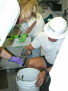
Scientists sift through sample buckets from an ROV dive. Click image for larger view.
Simply stated, multiple photographs are taken along sections of the structure or cell that you want to observe. For example, let's say you wish to study the effects of a bioactive compound on microtubules (a structural feature within the cell). Multiple microscopic images are made of the microtubule structure and stored in a database. Specialized software will take the two-dimensional pictures and put them together, forming a 3-D image that the researcher can view from any angle.
Another interesting feature of confocal microscopy is the use of fluorescence as a type of "tag" to examine cellular structures, unique microorganisms that might be living in the tissues of the organism, and the presence or absence of proteins within cells. Fluorescence is the giving off of light when an outside source of light is introduced. You see this with many types of paints that seem to glow when hit with "black lights." You can use fluorescent dyes to tag specific structures or proteins within a cell, and then observe them using the 3-D imagery of the confocal microscope.
The numerous samples collected during this cruise will be sent to HBOI, where they will undergo intensive research for months to come. Confocal microscopy is only one of the tools that Tara and her colleagues will employ to further investagate potential biomedical compounds that come from the sea.
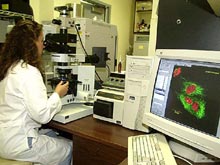
A typical confocal system consists of a high-quality fluorescent microscope, lasers, a control unit, photomultiplier tubes and mirrors, and a high-speed computer. Click image for larger view.
The Confocal System
Tara Pitts
Division of Biomedical Marine Research (DBMR)
Harbor Branch Oceanographic Institution
Confocal microscopy is a technique used to construct digital images of microscopic samples. This form of microscopy is typically used for examining samples that can be stained with fluorescent dyes, as well as samples that autofluoresce, such as the photosynthetic (use of sunlight to produce energy) pigments in most algae. It can also be used to examine specimens with transmitted light.
A typical confocal system consists of a high-quality fluorescent microscope, lasers, a control unit, photomultiplier tubes and mirrors, and a high-speed computer. The system requires a cool environment, a vibration-free platform, and an area that can be darkened. The confocal operator must have basic knowledge and skills in microscopy. The operator must also be trained to use the system and its specialized software.
The confocal system focuses on one plane of view at a time and records a digital image. All the planes are combined to form a focused three-dimensional image. Special software allows the user to view either the entire image or each focused plane. The advantage of using confocal microscopy over standard fluorescent microscopy is the feature that enables the scientist to examine cells or cell structures in three dimensions. With the older technology, only one plane of an image can be viewed at a time, while the rest of the image is often out of focus. Confocal microscopy allows one to see an entire specimen or parts of a specimen in a sharp 3-D image. After acquiring this digital image, the software allows the user to manipulate the image to improve its quality. The image can then be saved as a file and imported or exported for use in other software programs. It produces quality images for publications, posters, presentations and Web sites.
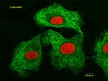
A fluorescent image of untreated cancer cells. Microtubules, which form the cells' cytoskeleton, are stained green. The cell nuclei are stained red. Click image for larger view.
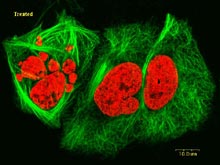
An image of trated cancer cells. These cells were exposed to a drug for 24 hours before being fixed and stained for viewing. The cytoskeletons are stained green; the nuclei are stained red. Click image for larger view.
Using confocal microscopy, scientists can study both living and preserved samples. One application for which confocal microscopy has been particularly useful is evaluating the effects of chemicals produced by marine organisms on specific structures or processes in cancer cells. This has helped scientists determine the mechanism by which these chemicals kill cancer cells, and could lead to the development of new drugs to treat cancer. Another application of this technology is the identification of unique microorganisms that live in marine invertebrates such as sponges.
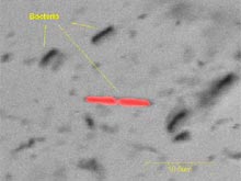
Bacteria from a sponge cell suspension showing some autofluorescent bacteria. Transmitted light and Krypton laser. Click image for larger view.
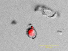
Autofluorescent microorganism from a sponge cell suspension. Transmitted light and Krypton laser. Click image for larger view.
In some cases, the bioactive chemicals may be produced by microbial symbionts, and confocal microscopy could be useful in identifying the type and location of the microbe in its host.
Sign up for the Ocean
Explorer E-mail Update List.