


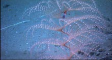
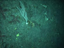
Lovely spiraling Iridigorgia coral, with brightly colored commensal shrimp. Click image for larger view.
Let's Go!
May 21, 2004
Mary Grady
Adjunct Earth Science Instructor
Northeastern University
Diana Payne
Biologist and Education Coordinator
Connecticut Sea Grant
It is to the sea that man must turn to meet the last great challenge of exploration this side of outer space.
—H. B. Stewart, Deep Challenge
Thursday afternoon, the education team broadcast their fourth day of live programs on the Internet. Each Webcast features a scientist describing what they are seeing on the remotely operated vehicle (ROV) cameras. Also included are one or two pre-taped segments on the topic of the day, such as how seamounts form or how the ROV technology works. Tolland High School teacher Lance Arnold has hosted each of the programs, and the scientists have taken turns providing the live voice-over.
Each program requires the cooperation and skills of a wide range of professionals. Thursday's Webcast, for example, involved the combined efforts of Institute for Exploration (IFE) ROV engineers Jim Newman, Todd Gregory, and Jim Miller; ROV camera operator Pamela Lezaeta-Sotomayor; and Dr. Scott France and Dr. Peter Auster as the "voice of science." Ivar Babb of the University of Connecticut ran the camera, while Connecticut Sea Grant education coordinator Diana Payne organized the scripts and hosted the live chat feature on www.explorethesea.com ![]() . Catalina Martinez, expedition coordinator from NOAA's Office of Ocean Exploration, acted as director, transmitting cues from the video crew to the on-screen "talent." The crew of the Ronald H. Brown were as helpful as ever, responding immediately to every request to move the ship to accommodate a shot.
. Catalina Martinez, expedition coordinator from NOAA's Office of Ocean Exploration, acted as director, transmitting cues from the video crew to the on-screen "talent." The crew of the Ronald H. Brown were as helpful as ever, responding immediately to every request to move the ship to accommodate a shot.
The science team continues to collect specimens and to see things never seen before. They accumulate information not only from the samples brought up in the bio box, but also from observing creatures in their natural habitat on the video monitors during dives, and from reviewing tapes afterwards. During Thursday's dive, they saw several Iridigorgia spiral corals harboring colorful shrimp that were full of eggs. "It’s been amazing to witness the organisms associated with the corals on this trip, as most scientists never have this opportunity," commented Dr. Watling. "You can’t possibly determine which organisms are associated with specific coral species by dragging a net over the bottom. You really need to observe the corals prior to collecting them."
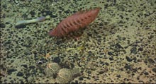
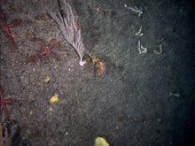
The scientists were also able to witness crab behavior, including a crab eating a shrimp, a male red crab "guarding" a female crab, and a crab aggressively protecting prime habitat.
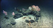
A crab strikes an aggressive pose to protect this oasis of spectacular diversity. Click image for larger view.
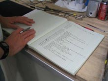
Even in the age of computers, scientists find handwritten notes invaluable. This is Dr. Jon Moore's notebook. Click image for larger view.
Today, the seas picked up for the first time on this trip. Swells and whitecaps followed everywhere we went. In the lab, chairs slid back and forth across the floor. Argus, tethered to the ship, was jerked up and down as the ship rode the swells. It created a challenge for the ROV pilots, but it didn't prevent them from continuing the dive, which went on for about 14 hours. By the time the dive ended mid-afternoon, the bio boxes were full. There was concern that it might be difficult to get the ROVs back on board in the ocean swells, but the operation was completed safely, and the science team was ready with their buckets to offload the samples.
On a research trip like this, the sheer volume of data collected is enormous. Every second of videotape shot during the ROV dives is saved for future analysis and reference. Still-shots and video clips are culled from the many hours of tapes. In the lab, specimens are examined under microscopes that have video cameras attached, still-shots are collected, and the film is archived. Many of the corals are laid out on a black background and photographed. Then tiny pieces from each coral are snipped off and stockpiled in sealed vials, to be analyzed back on shore. Handwritten lists of specimens and where they were found are meticulously recorded. The collection is carefully doled out to university labs and museums. Nothing goes to waste.
Late Thursday night, after much discussion about the various options available and the remaining objectives of all on board, the decision was made to steam overnight to Balanus Seamount and dive there Friday morning. This seamount has been explored only once before—30 years ago—by Bob Ballard and colleagues in the Alvin submersible.
Overnight, the SeaBeam multibeam sonar crew acquired data and found that the bathymetry of Balanus Seamount was different than they had expected. The mountain has three distinct peaks. The science crew used that image early Friday morning to choose the dive site along a steep ridge.
Friday morning dawned with more calm seas and bright sun. The ROVs went back into the water, and by mid-morning they were sending back video. The Webcast team set up for their last live program of the week, which included a segment about the value of art in underwater landscape interpretation. All day and all night long the exploration continued, proving once again the utility of Hercules and Argus for scientific research.
During the day, the science lab cleared out quickly when a call came over the intercom that the bridge crew had spotted a mola mola fish off the port stern. A half-dozen scientists ran out to look. The big mola mola, also known as an ocean sunfish, was swimming close to the surface, even breaking slightly above the waves, right below the rail.
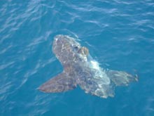
A mola mola soaks up the sun. Click image for larger view.
Catalina Martinez and Les Watling inspect their sargassum catch. Click image for larger view.
Later in the day, Catalina Martinez, expedition coordinator for NOAA's Office of Ocean Exploration, noticed sargassum seaweed, with tiny fish schooling around it, drifting near the ship. With help from Dr. Les Watling, she dropped a bucket overboard, drew it up full of seawater, and retrieved a small mat of sargassum. No fish were caught, but a tiny shrimp and a delicate nudibranch (marine slug) were found nestled within the sargassum. A group gathered to watch the elegant way the nudibranch undulated through the water. After a few minutes of observation, the sargassum with its inhabitants was gently placed back into the sea.
It took the coordinated efforts of two ROV pilots to collect fossil corals with a scoop-action, using a net and the Hercules scissor claw. Click image for larger view.
For most of the day, Hercules roamed the slopes of Balanus, photo-documenting and collecting specimens. ROV pilot Tom Orvosh practiced using the manipulator arm. ROV pilot Dave Wright talked him through, with suggestions on the complex strategy and technique required to collect the fragile corals and get them safely stored in the bio box. A piece of rope, tangled into some corals, came into view on the video monitor. This was one of the few signs of manmade debris we saw at depth on this trip.
During dives, the science teams often make decisions on the fly about which direction to take the ROVs. Much depends on what they are seeing from moment to moment. At times, it comes down to the classic words that have probably been said by explorers throughout history: "Let's go and see what's there."
At one point during the dive on Balanus, the ROV pilots collected fossil corals using a net and the Hercules scissor claw. It was a challenge, trying to coordinate the movements of the two arms, but through the skilled work of ROV pilots Tom Orvosh and Dave Wright, a sample was successfully collected.
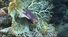
A bouquet of Corallium with deep purple Trachythela octocoral, brittle stars, crinoids, and sponges. Click image for larger view.
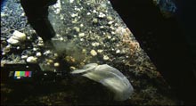
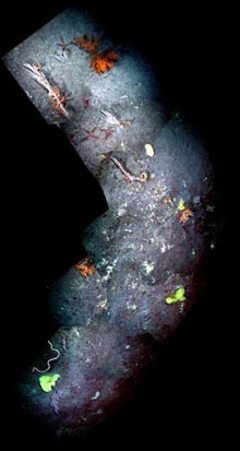
A phenomenal line-up of organisms representing the great diversity on Balanus Seamount: a strange spoon worm, an elegant sea pen, a stalked crinoid, and two xenophyophores with brittle stars. Click image for larger view.
After the dive, Dr. Les Watling surfed through high resolution still images. He commented that he had never seen so much Corallium coral in one place before, and that the diversity of Balanus was spectacular.
This image (Figure X) shows a sample raw image, which shows only a single coral and doesn’t accurately represent the colors and available light. Click image for larger view.
Photomosaicing
Allan Gontz
Graduate Student
University of Maine
Jonathan Howland
Senior Engineer
Woods Hole Oceanographic Institution
Adjunct Engineer, Institute for Exploration
Scientists use a variety of mapping techniques to better understand the sea floor and its inhabitants. One of the tasks frequently performed in undersea mapping is "photomosaicing." Photomosaicing is an imaging technique that overcomes the limited field of view of cameras by combining multiple, overlapping individual images into a larger composite picture, or photomosaic. Surveyors and scientists have been using mosaicing in terrestrial applications for decades. Within the past ten years, primarily due to the increasing availability of digital cameras and inexpensive desktop computing, photomosaicing techniques have shifted from film and darkroom manipulation to purely digital techniques. In recent years, it has become common for digital-camera vendors to include photomosaicing software as part of their consumer-oriented products (although they frequently call it “panorama” software).
Photomosaicing is particularly useful for underwater applications, since the water drastically limits the range and field of view of optical sensors. Oceanographers began making photomosaics as soon as underwater photography became practical. The deep-sea environment presents many challenges to typical photomosaicing tools, including varying illumination, severed terrain-induced parallax (the relative change in positions of objects in images caused by movement of the camera), and unstructured motion of the camera. These physical constraints have limited the applicability of the terrestrial tools to underwater problems.
As an example of the photomosaicing process, consider the following: The Hercules ROV carries two digital still cameras, one black and white and one color, which collect a new image every 10 seconds. Some of the biologists on board the Ronald H. Brown wanted a larger view of a field of coral so that they could better understand the relative positions of the individual corals. Color imagery from the period of interest was retrieved from the on-line archive. Figure X shows a sample raw image, which shows only a single coral, and which doesn’t accurately represent the colors and available light.
This is the same image as Figure X after color balancing and other image enhancement operations. Click image for larger view.
Different frequencies (colors) of light attenuate (fade) at different rates in the water, so underwater imagery frequently doesn’t look “right.” Red is attenuated most quickly, so underwater imagery is frequently dominated by blue and green. A process known as “color balancing” can restore a more natural range of colors to the imagery. The colors can be controlled or verified by examination of well-lit close- up imagery or of samples physically retrieved from the sea floor. Figure Y shows the same image after color balancing and other image-enhancement operations.
These operations were carried out on all of the selected imagery. A single central image was selected as a base for the photomosaic. Successive images were compared to the base or to the growing mosaic. Features that could be identified as common between the candidate image and the mosaic were measured, and the candidate images were warped so that the features would match when the images were digitally combined. A digital “cut-line” was marked in the image overlap area, and the candidate image was blended into the mosaic.
This image is the final mosaic given to the science party. Click image for larger view.
By repeating this technique with many images, a photomosaic is built up. Because a two-dimensional mosaic cannot accurately represent irregularly shaped terrain, errors grow throughout the process, and the area coverage of the final mosaic is frequently limited by this error growth. Figure Z shows the final mosaic that was given to the science party.
As previously stated, automatic mosaicing is relatively common in terrestrial practice. It is an active research topic in underwater application. As automatic mosaicing technology matures, it will free the mosaicer from the tedious task of marking overlap points. Rigorous mathematical modeling will more accurately represent the terrain, allowing larger mosaics, as well as derivation of the shape of the terrain from the images themselves.
Sign up for the Ocean Explorer E-mail Update List.